Elastic Veterinary Kinesiology Taping (K-Taping)
The Kinesio Taping® Method is a rehabilitative technique designed to facilitate the body’s natural healing process by providing extended soft tissue manipulation. This manipulation mimics the effects of massage while at the same time providing support and stability to muscles and joints without restricting their range of motion.
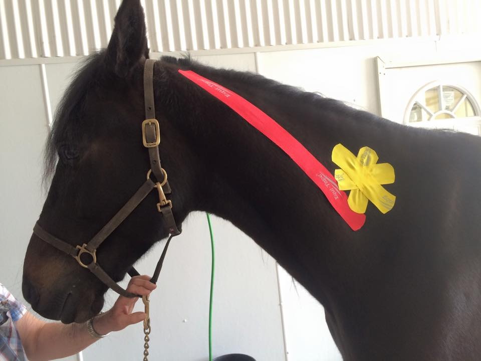
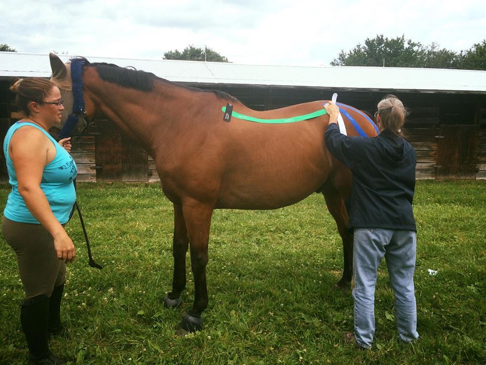
Kinesiology taping to relax hypertonic neck muscles (red) and activate an acupuncture point (yellow) prior to a chiropractic adjustment
The Kinesio Taping method was developed by Dr. Kenzo Kase in 1973 to provide a non-pharmaceutical method to alleviate his patients’ pain in between appointments. He created the tape with a thickness, stretch, and weight similar to the superficial skin layer. This allows the tapes’ effects to target different receptors within the somatosensory system with minimal perception of its presence on the skin. The tape ‘breathes’ allowing for water evaporation and cooling. The adhesive pattern is in a repeated wave form, similar to a fingerprint. This pattern helps lift the skin and allow moisture to escape. The tapes’ elastic quality causes tissue stretching that creates either a massage or a lifting effect on the sub dermal and fascial layers. This forms convolutions in the skin, whether visible or microscopic, which separate the muscle and dermal layers as well as increase interstitial space and stimulate mechanoreceptors. The increased space has a lower pressure gradient than the surrounding areas and pulls fluid and exudates into it. This fluid movement essentially decreases inflammation (heat, pain and swelling) in the treated area and aids in normal blood flow and lymphatic drainage. The space also takes pressure off swollen or injured muscles, reducing pain and allowing tissue remodeling and healing. In addition, the tapes massaging effect provides stimulation to skin cells that affect pain pathways—similar to rubbing a spot that hurts.
Kinesiotape comes in several colors. The colors do not have different properties. However, colors on the darker end of the light spectrum (red, black) will absorb more sunlight and increase skin temperature under the tape. If heat is a desired part of the treatment red or black tape may be incorporated. If inflammation is present, ie tendonitis, a ‘cooler’ color can be used. This effect is more drastic in humans than horses due to the horses' coat.
Kinesiotaping can be used at any point in the healing process, from acute onset to sub acute, chronic, or into the rehabilitative stage. Kinesiotaping may also be used as a preventative modality in the face of rehabilitation or strenuous exercise. Its ability to re-educate the neuromuscular system, optimize performance, prevent injury, promote good circulation, and reduce the neuromuscular stresses of exercise, makes it perfect as a preventative therapy. It can be used in conjunction with cryotherapy, hydrotherapy, acupuncture, chiropractic treatment and during workouts. It should not be used in infected areas or areas with known tumors or directly over open or healing wounds. Deep vein thrombosis should also not be taped.
When used properly, Kinesiotaping provides support during movement, improves circulation and lymphatic flow, reduces pain, inflammation and tension, relaxes and assists muscles, releases fascia, and improves joint function. These all result in faster and more complete healing and improved movement and positioning.
Kinesiotaping has been proven to have positive physiological effects on the dermal (skin), lymphatic, circulatory, and neuromuscular (fascia, muscles, ligaments, tendons, and joints) systems. Kinesiotaping influences these systems through four major actions:
Supports weakened, over used or injured muscles
by assisting the muscle in either contraction or relaxation- depending upon the tape application
by reducing muscle fatigue and cramping
by preventing over extension and over contraction
by reducing fatigue and preventing re-injury
Improves circulation (lymphatic and blood flow) & removal of congestion
by lifting dermal and fascial layers to create interstitial space
by creating osmotic pressure gradients that cause fluid flow
by creating drainage pathways in the subdermal layers
by massaging fluid along theses created pathways
which reduces inflammation (heat, pain, swelling), removes lactic acid, break down products, damaged cells and cellular debris, and brings in the components required for healing
Reduces pain
by relieving tension in hypertonic, over used or cramped muscles
by improving circulation which reduces the swelling, removing the pressure that stimulates the pain pathways
which facilities normal motion and prevents secondary dis- or mis-use injuries
Corrects joint problems
by stabilizing the ligaments and tendons that support the joint
by allowing full range of motion, thereby preventing adhesion formation during healing that result in a limited or frozen joint
by reducing joint swelling
by facilitating proper positioning, use and motion of the involved structures during healing.
These four actions can be utilized in many variations and combinations in order to achieve the treatment goal; desired physiological correction. The most common corrections utilized include: circulatory, fascial, functional, ligament or tendon, mechanical, scar tissue, or spacial correction.
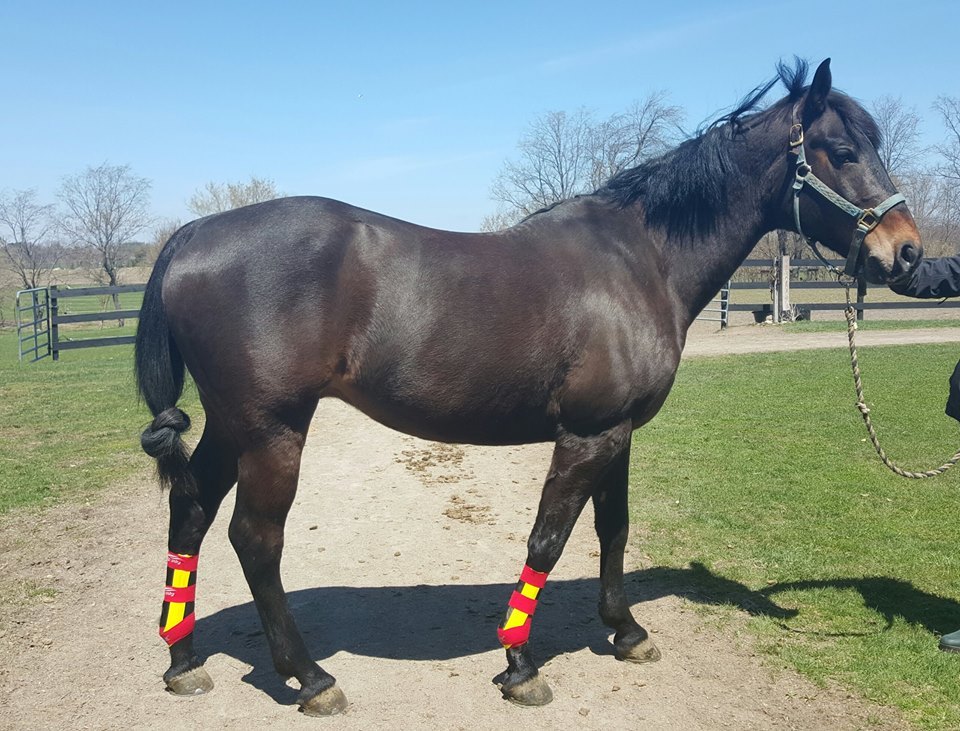
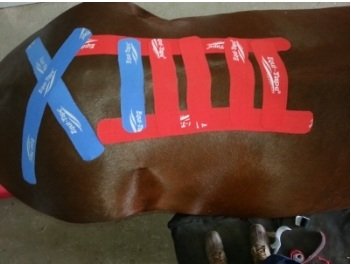
Belgian gelding treated for a "Boggy Hock"
Circulatory / Lymphatic Correction
This application enhances fluid, congestion, exudate, and blood flow between tissue layers; reduces edema and drops the temperature of the inflamed tissue, as well as reduces pain and discomfort and promotes healing. The tape is applied to swollen areas as overlapping fan strips. The tape create areas of decreased pressure that act as channels to direct the flow of exudates and extracellular fluid towards lymphatic ducts, facilitating their removal. The overlapping tape tails exert a multi directional pull in the soft tissues every time the horse moves. This encourages the opening and closing of pre-lymphatic collector and angion vessels that gather interstitial fluid (outside the vessels, between the cells). It also pulls/guides exudates through the superficial tissues to less congested areas. This reduction in fluid also reduces the inflammatory and edematous pain. The tape works as a massaging pump to move the exudates out of the area and allow more congestion (interstitial fluid) to fill the collecting vessels and in turn be moved up the lymphatic ducts and back into the blood stream.
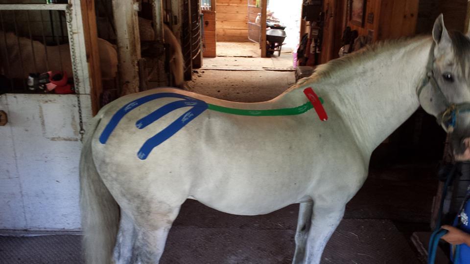
Fascia Correction
This application reduces tension or adhesions between and within the fascia layers that are limiting motion. The tape is applied with zones of varying tension to create convolutions that stimulate, through the skin and tape motion, a micro-massage effect on the fascial tissue. The depth and severity of an adhesion may be targeted by changing the direction and force of the convolutions by applying the differing tape widths with a high or low pressure or tension.
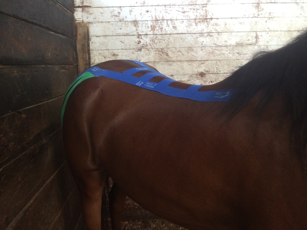
The green tape is performing a Fascial Correction on the neck. The red and blue "star" is treating an acupuncture point.
In order to fully understand the effects of fascial correction, the fascial tissue must be understood. Fascia is the dense connective tissue covering that separates muscle bodies and provides mechanical support and frame work to other tissues of the body. The mechanical tension generated by muscle activity is transmitted through the fascia as proprioceptive stimuli to the brain. It tells the body where its parts are at all times without having to see them in order to regulate posture and movement. Fascia is comprised predominantly of type I connective tissue in a latticed pattern but also contains elastic fibers and contractile cells. The presence of these cells suggest fascia may also influence musculoskeletal dynamics through a smooth-muscle like contraction. Superficial fascia is found under the skin and extends all over our body from head to toe. Some researchers consider it to be the communication network of our bodies. Deep fascia is the envelope covering each muscle. In essence every muscle fiber is wrapped with fascia as well as the whole muscle and then the entire body. Between the superficial and deep fascia there is a continuity of fascial and fascial planes throughout our body. This fascial movement throughout the body provides proprioceptive feedback to the brain and body. You know when you are leaning back because the fascial is sending stimulus to your brain about the position of your muscles and body parts. Fascia allows the continuous transfer of information about location and tension at all times everywhere in the musculoskeletal system. Therefore, flawless effortless motion requires flawless uninterrupted unimpeded movement throughout the fascial planes. Any adhesion, scar, or limitation changes that information highway. Research articles have shown the majority of musculoskeletal injuries are dysfunctions of the superficial and deep fascia.
Functional Correction
This application will either assist or restrict a motion (flexion or extension) by changing the brains perception of the joints’ position thru increased tension at the skin or joint capsule. The tape is placed under tension while the joint is at the end of its range of motion, either flexion or extension. This creates space over the muscle, tendon, or joint area being treated when the joint is in its desired position, and stimulates mechanoreceptors in the skin and joint capsule when the joint is nearing the end of its range of motion. The same tension that triggers these proprioceptive impulses also acts like a pre-load on the tendon, muscle, or joint; mimics the tension in the structure just before it reaches its end range of motion, warning of imminent injury if it moves further. These two actions cause the brain to send out nerve signals to stimulate the opposite movement. This means that the tape will assist motion of a muscle or joint (either contraction of extension) and resist the opposite motion.
For example, if a joint needs assistance in flexion and resisting extension, the tape would be applied so that during extension the tension produced by the tape on the skin increases to a level where the resulting stimulus is perceived as the joint having reached the end of normal joint motion. This causes the body to limit overstretching or hyper-mobility of the injured structure and therefore re-injury. Then flexion will be assisted by the same increased tension, which was perceived as end range of motion tension and signals a return of the structure to the desired position. This is the point where the tape tension lifts the skin, creating space below the skin. The space reduces the mechanoreceptor stimulation triggered by the tape when the joint is either flexed or extended (depending upon the tape application).
To accomplish all these activities, the tape must be applied under tension so that the action of flexion or extension causes a pull that will trigger skin and joint capsule mechanoreceptors to release stimuli to the brain. In response to the stimuli the brain will send impulses to muscles to either assist or limit a joint motion. In essence, the body will change joint position to return to normal the increased tension on the skin. The use of kinesiotape in a Functional correction capacity is complicated but will stabilize a joint when the supporting ligamentous structures have been injured, without preventing joint motion which can cause adhesions.
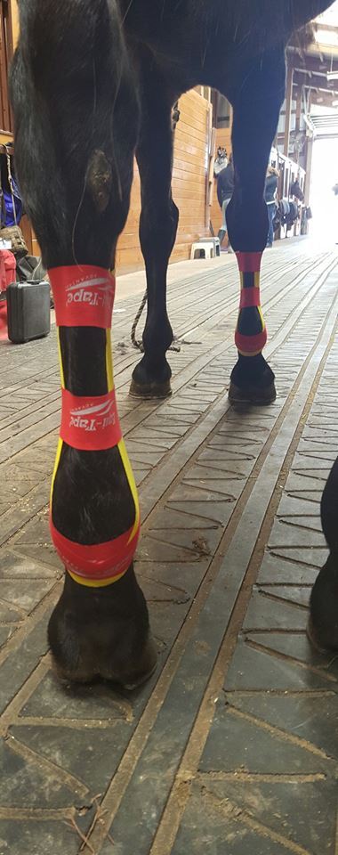
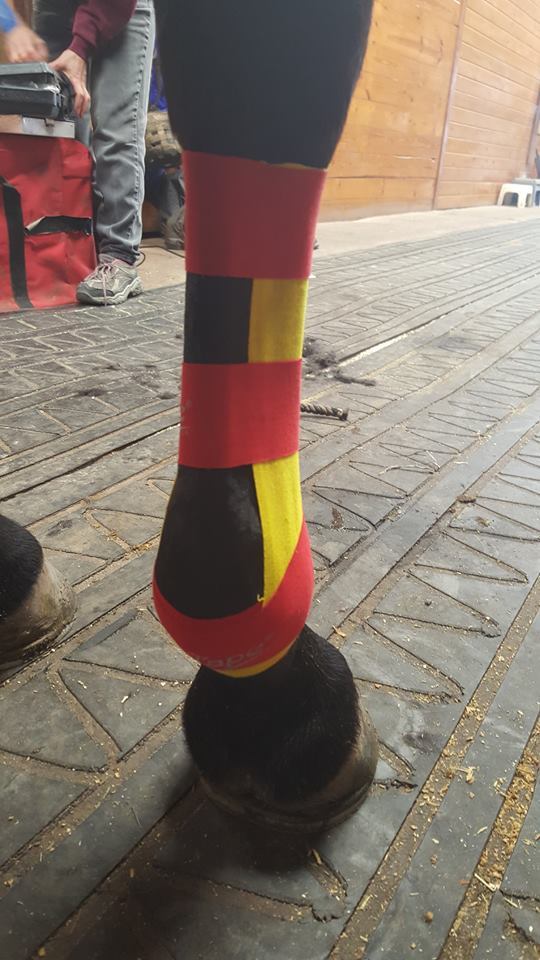
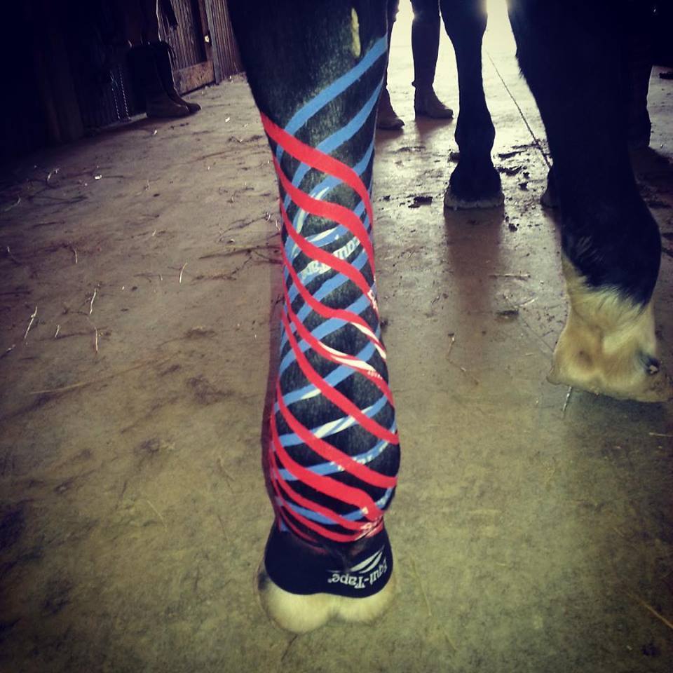
Left Hind taped with a polo wrap for heat activation of the adhesive for taping longevity. Right hind demonstrates taping for support of Suspensory apparatus
Ligament/tendon correction
This application supports injured tendons and ligaments without limiting motion in order to facilitate proper realignment of strained or torn fibers during healing without over use and reinjury. The tape is applied over an injured tendon or ligament to stimulate the mechanoreceptors in the skin or joint capsule and influence the golgi tendon organ. This organ is located within every muscle spindle and is the primary receptor transmitting information to the brain about the specific muscle tissues’ tension level (how relaxed or contracted it is). The brain receives these stimuli as proprioceptive stimuli. The goal of the tape is to create mechanoreceptor and golgi tendon organ input similar to the pre injury state. In response to the healthy state signals, the brain sends impulses for normal, not protective motion and use of the structure. In order to support the injured structure during this motion, the tape is applied with more stretch than other applications. Depending upon the severity of the injury, other motion limiting wraps may be required in addition to Kinesiotaping. The tape may be used simultaneously with poultices, sweats and medicated wraps without inhibiting their effects.
Mechanical Correction
This application provides functional support to muscles, fascial tissue or joints without preventing active range of motion or inhibiting the circulation. The tape may be applied to either directly assist in the correct anatomical positioning of the structure or to prevent pathological motion. The correct positioning is attained by stimulating a sensation in the skin, in multiple or deeper muscles, or in multiple tissue layers which causes the body to adapt to the stimulus. The amount of stretch the tape is applied with as well as the downward pressure, determines the depth and perception of skin movement, providing a positional stimulus to the mechanoreceptors. Their feedback to the brain triggers impulses to adjust the structures position in order to minimize mechanoreceptor stimulation by the tapes tension. Similar to its use in ligament and tendon correction, the tape is used to maintain the structure in a desired position or range of motion. In like manner the tape may be applied to have increased tension and therefore mechanoreceptor stimulation when the structure is nearing the end of its healing range of motion. In this case the impulses released by the brain stop the structure from moving further in that direction, effecting a ‘blocking action’ of joint or tissue motion to prevent pathological motion. Thus in this technique the tape is used to stimulate the mechanoreceptors which then regulate the bodies positioning of the muscle, fascia, and joint position, either blocking or stimulating muscles and motion.
Scar Tissue Correction
This application is used to reorganize and minimize scars and scar tissue. The tape is applied in two phases. The first phase is applied in repetitive strips that stimulate mechanoreceptors to send stimuli that facilitates both relaxation and contraction (opposing forces) in adjacent lines. It works like cross friction massage to disrupt and disorganize the scar. This phase may be reapplied for one to several months depending upon the scar size and age, how old it is. Once the scar has ‘softened’ the second phase begins. The tape is applied to create forces in the same plane in order to facilitate reorganization and realignment of the scar tissue. When scar fibers are in alignment the scar size is smaller and the surrounding tissue is less compromised by tension at the scar interface.
Space Correction
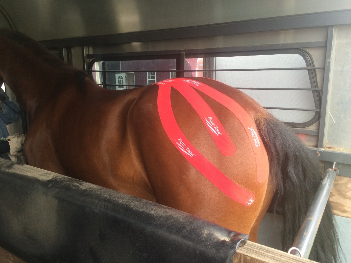
Space correction to relieve inflammation after a shoulder contusion
This application is used to decrease pressure and pain. The tape is applied in a manner that emphasizes its elastic qualities and lifting effect in order to ‘lift’ the skin, fascial and soft tissue over a target area. This slight lifting creates space directly above the area of pain, inflammation, swelling or edema. The increased space decreases the pressure in that area, reducing the amount of irritation on the chemical and nociceptors (pain receptors, thereby decreasing the pain. The increased space also allows for lymphatic and circulatory vessels to fill easier and speed the removal of inflammatory fluid, cellular exudates and break down products from the injury. The mechanoreceptors directly stimulated by the tape also trigger the gate control theory of pain and drop the pain levels. Just like walking off a stubbed toe, the motion nerves ‘shout’ louder than the pain nerves and ‘drown out’ the pain sensation. The tape can be applied to lift a focal area, an area around a focal site, or to lift a larger area and facilitate drainage through channels in a certain direction.
Enjoy our gallery to see the wide variety of applications of K-Taping!